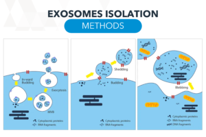
It is critical to understand the importance of exosome isolation methods, which play key roles in cell-to-cell communication and disease mechanisms. Exosomes are tiny vesicles released by cells that contain biomolecules such as proteins and sequences that can affect the functions of surrounding cells. By understanding how exosomes are isolated, we can study the mechanism of cell-to-cell message transmission, thereby revealing the occurrence and development of many diseases.
The study of exosomes is not only of great significance to basic science, but also expected. Therefore, an in-depth understanding of exosome isolation methods will not only help reveal the mysteries of life, but also has the potential to promote innovation and progress in the medical field.
It is worth noting that since the biological origin of various vesicles is difficult to clearly identify, the International Society for Extracellular Vesicles (ISEV) recommended in the MISEV2018 guideline that they be named in the following way:
It is worth noting that since the biological origin of various vesicles is difficult to clearly identify, the International Society for Extracellular Vesicles (ISEV) recommended in the MISEV2018 guideline that they be named in the following way:
Naming basis | Physical characteristics | Surface markers; biochemical composition | Descriptions of conditions or cell of origin |
|---|---|---|---|
Example | •Exosome Size: small / medium / large extracellular vesicles (sEVs / mEVs / lEVs) | •D63+ / CD81+ EVs | • Podocyte EVs |
•Density: slow / middle, high EVs | •Annexin A5-stained EVs | • Hypoxic EVs | |
• Large oncosomes | |||
• Apoptotic bodies |

Compared with synthetic carriers such as liposomes and nanoparticles, exosomes have characteristics such as endogeneity and heterogeneity, making exosomes useful as Excellent carriers can transport bioactive substances to target cells through a variety of pathways and sites to participate in regulation, such as tissue repair, Immune regulation, angiogenesis, cellular differentiation, neoplasia, etc.
Therefore, exosomes have great potential and advantages in disease diagnosis, treatment and biology research, such as as biomarkers for tumor diagnosis and drugs for cancer treatment. carrier etc. However, exosomes also have some limitations, such as low stability, low yield, low purity, and weak targeting, which may limit their clinical application.
Extracellular vesicles, EVs | Exosomes | Microvesicles | Apoptotic bodies |
|---|---|---|---|
Size
| 40~120 nm
| 100~1000 nm
| 1000~5000 nm
|
Biogenesis | Multivesicular bodies, MVB
Endo-lysosomal pathway | Plasma membrane, outward budding | Plasma membrane, apoptosis process |
As the research on exosomes isolation continues to deepen, their potential application value is also continuously being explored. The isolation, purification, and concentration (also known as enrichment) of exosomes are critical for evaluating their biological functions and their downstream applications. However, the components of biological samples are complex and contain many similar molecular structures, such as cell fragments, protein aggregates, lipoproteins, etc., and the size and composition of exosomes are heterogeneous, making “isolation” more challenging. Therefore, “how to effectively isolate and concentrate exosomes” is a major test faced in academic research and clinical applications.
There are many exosomes isolation methods, depending on the principles. Common exosomes isolation methods are ultracentrifugation, particle size screening chromatography, ultrafiltration, precipitation and immunoaffinity. The following table summarizes the differences between the methods:
Ultracentrifugation (UC) *1 | Particle size screening chromatography (SEC) | Ultrafiltration (UF) | Precipitation | Immunoaffinity (IA) | |
|---|---|---|---|---|---|
Principle | Sedimentation Coefficient (size, density)
| Hydrodynamic Radius (size, molecular weight)
| Membrane pore size (size, molecular weight)
| Solubility (surface charge)
| Specific Binding (Membrane protein marker)
|
Yield | Low | Medium | High | High | Low
|
Purity | Medium (lipoprotein)
| Medium to high (lipoprotein, albumin)
| Medium (protein)
| Low (proteins, polymers)
| High
|
Functionality | Medium
| High | Medium | Low
| Low
|
Time | > 4 hr
| 0.3 hr*2
| < 4 hr
| ≈0.3~12 hr
| 4~20 hr
|
Sample volume
| Large | Medium | Large *(TFF)
| Large | Small |
Denature and inactive of EVs
| Yes (high speed)
| No | Yes (Shear force)
| Yes (Co-precipitates with proteins or polymers)
| Yes (Refining steps)
|
Complexity | Medium
| Simple | Simple | Simple | Medium |
Scalability | Medium
| High | Medium to high 3(TFF)
| High | Low |
Other | • Labor intensive and time consuming | • High reproducibility
• EVs have complete functions and forms | • High reproducibility
• High flexibility of use
• Filter membrane clogged | • Polymers are difficult to separate
• More costly kit | • EVs have the best purity
• Study of specific EV subpopulations
• Unable to separate total EVs
• Antibodies are expensive |
*1 Ultracentrifugation (UC) is also called differential centrifugation (dUC)
*2 EVs purification can be completed within 20 minutes using commercially available kits
*3 High scalability refers to TFF tangential flow filtration method
Ultrafiltration (UF) is a method of separation based on molecular size, which can be subdivided into centrifugal-driven ultrafiltration centrifuge tubes, pressure-driven stirred filters, and tangential flow filtration (TFF). type. Ultrafiltration centrifugal tubes and stirred filters are easy to operate and have fast processing time, but the biggest problem is that the filter membrane is easily clogged, which not only reduces the separation efficiency but also increases the cost; and the development of tangential flow filtration technology (TFF) It can significantly solve the problem of filter membrane clogging.

Many documents also point out that when TFF is used to separate EVs, its yield, reproducibility, and decontamination capabilities are superior to the ultracentrifugation method (UC or dUC) called the gold standard for EV purification, and even saves more than 40% of time. , more suitable for large-scale mass production applications.
For a detailed comparison of each method and the EV isolation data of TFF & UC, you can watch the “Exosome Exosome Purification Online Seminar Live Highlights”.

*LSC:Low speed centrifugation

We equip your laboratory with speed and efficiency
Fill in the form and our team of experts will recommend the best viscometer and setup based on your requirements.